Sunday, September 30, 2007

Friday, August 17, 2007

PALMER'S POINT

In an illustration on page 2 chapter 1 , Palmer's point is shown, however; no explanation is given.

The following article is discussing the minimally invasive approaches through the abdominal wall in laparoscopy and you can find that in case of Central adhesion around the umbilicus, the Palmer's
point is considered to be the first alternative root to enter the laparoscopy port.

Article 1

URACHUS AND VITELLINE DUCT


The
urachus is an embryological canal connecting the urinary bladder of the fetus with the allantois a structure that contributes to the formation of the umbilical cord. The lumen (inside) of the urachus is normally obliterated during embryonic development, transforming the urachus into a solid cord, a functionless remnant.


Allantois

This sac-like structure is primarily involved in respiration and excretion, and is webbed with blood vessles.
The function of the allantois is to collect liquid waste from the embryo, as well as to exchange gases used by the embryo.
In placental mammals, the allantois is part of and forms an axis for the development of the umbilical cord.


Vitelline duct

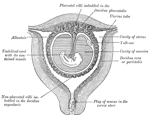
At the end of the fourth week the yolk-sac presents the appearance of a small pear-shaped vesicle (umbilical vesicle) opening into the digestive tube by a long narrow tube, the vitelline duct.
The vesicle can be seen in the after birth as a small, somewhat oval-shaped body whose diameter varies from 1 mm. to 5 mm, it is situated between the amnion and the chorion and may lie on or at a varying distance from the placenta.

As a rule, the duct undergoes complete obliteration during the seventh week, but in about three per cent of cases its proximal part persists as a diverticulum from the small intestine,
Meckel's diverticulum, which is situated about two feet above the ileocolic junction, and may be attached by a fibrous cord to the abdominal wall at the umbilicus.
Sometimes a narrowing of the lumen of the ileus is seen opposite the site of attachment of the duct.


UMBILICUS

The umbilicus is essentially a scar made from the remnants of the umbilical cord.
It is situated in the Linea Alba and in a variable position depending on the obesity of the patient.

however, the base of the umbilicus is the thinnest part of the abdominal wall and is the commonest site of insertion of the primary port in laparoscopy.

LINEA ALBA & PYRAMIDALIS MUSCLE

Where the abdominal muscles coalesce in the midline, linea alba is formed.
Pyramidalis muscle is present in all women origination on the anterior surface of the pubis and inserting into the linea alba.


Monday, August 13, 2007

The anterior abdominal wall


MAYLARD INCISION OF THE ABDOMINAL WALL

Cares need to be taken to avoid the inferior epigasteric vessels while using accessory trochars during laparascopy and to ensure that they are identified and closed appropriately when making a maylard incision of the abdominal wall.

Pfannenstiel incision

The Pfannenstiel incision results in good exposure to the central pelvis but limits exposure to the lateral pelvis and upper abdomen. These factors limit the usefulness of this incision for gynecologic cancer surgery. If the patient is thin and has a gynecoid or platypelloid pelvis, this incision can be used for a radical hysterectomy and pelvic lymph node dissection.

The incision is usually made 1-2 fingerbreadths above the pubic crest. Use of a marking pen is helpful to keep the incision symmetric. An incision length of 10-14 cm is sufficient. Increasing the length of the skin incision usually does not improve exposure due to the rectus muscles. The incision is made through the subcutaneous fat to the fascia. The superficial epigastric vessels are often near the lateral edges of the incision.

The anterior fascia is incised in the midline with a scalpel or electrocautery. Using curved scissors or electrocautery, the fascia is incised in a curvilinear fashion 1-2 cm lateral to the rectus muscle. The upper edge of the fascia is grasped with 2 Kocher clamps on either side of the midline. Using electrocautery, the rectus muscle is dissected free from the fascia. Electrocautery allows coagulation of multiple small vessels that perforate the rectus muscle to the fascia. The rectus muscles are mobilized off the fascia to the level of the umbilicus. Next, the lower fascial edge is grasped with Kocher clamps. Electrocautery is used again to dissect the rectus muscles and the pyramidalis muscle from the fascia. The rectus muscles are separated. The peritoneum is opened and incised vertically to complete a Pfannenstiel incision.

Closure of the Pfannenstiel incision is straightforward. The peritoneum does not need to be closed separately as re-epithelization occurs within 48 hours. Closure of the peritoneum does not add to the strength of the incision. Regardless of whether the peritoneum is closed, the rectus muscles should be thoroughly irrigated with water or saline, and any bleeding areas should be cauterized or ligated. Bleeding from small perforating vessels through the rectus muscle is the most common source of subfascial hematoma. The fascia is approximated with a delayed absorbable suture. Usually, a separate suture is started at each end of the fascial incision, and all layers of the anterior rectus sheath are incorporated. Unless a large area of dead space exists between the fascia and the skin, closure of the Scarpa fascia is not needed. Placement of a closed drainage system, like a Jackson-Pratt drain, may be needed if a large amount of fluid collection is anticipated.

Maylard incision

In an effort to improve surgical exposure to the lateral pelvic sidewall with a transverse incision, Maylard proposed a transverse muscle-splitting incision. This incision usually refers to a subumbilical transverse incision. For gynecologic surgery, the incision is made 3-8 cm superior to the pubis symphysis. The anterior rectus sheath is cut transversely. The inferior epigastric vessels are identified under the lateral edge of each rectus muscle and then are ligated. Patients with significant peripheral arterial disease may experience ischemia from ligation of the inferior epigastric vessels. These patients may have collateral flow from the epigastric vessels to the lower extremities. After ligation of the inferior epigastric vessels, electrocautery is used to transversely cut the rectus muscle. The peritoneum is opened and cut laterally.

To facilitate closure of a Maylard incision, flex the operating table. Close the peritoneum with an absorbable suture. Next, inspect the ties placed on each inferior epigastric vessel, and irrigate with water. Examine the cut edges of the rectus muscles for any bleeding areas. The fascia and underlying rectus muscle can be closed with a monofilament absorbable suture.